Mrs ANF, a 35 year old lay in her fifth pregnancy, presented with headache, nausea and vomiting and her period delayed 1 week. Pregnancy was suspected and she had come for a confirmatory ultrasound scan. There was no bleeding and she was otherwise well. Nothing unremarkable was found on physical examination. However, ultrasound scan showed a mass in the uterus which contained multiple small cystic structures, as seen here.
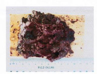
This was characteristic of molar pregnancy. She underwent suction curettage the next day and a large amount of tissue was removed.
This was confirmed as hydatidiform mole. She is now being monitored for recurrence and counselled to practice contraception for at least the next 6 months.
Hydatidiform mole or molar pregnancy is a rare growth that can occur when a woman gets pregnant. In most instances there is no fetus but occasionally a mole can occur together. Apart from being considered a miscarriage and cause bleeding, a molar pregnancy has the potential to spread out of the uterus and even turn cancerous. Thus steps must be taken to evacuate it completely and prevent progression to cancer.


No comments:
Post a Comment